

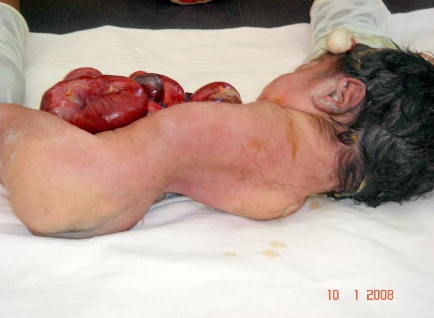
Omphalocele is a midline abdominal wall defect (absent skin, fascia & abdominal muscles) of variable size at the base of the umbilicus. The defect is covered by a three-layer membranous sac consisting of amnion, Wharton’s jelly and peritoneum.
The umbilical cord inserts at the apex of the sac. The sac contains herniated abdominal contents, usually bowel but may also have the liver, bladder, testis or ovary. They are generally classified as liver-containing and non-liver-containing.
Risk Factors
- Women in extremes of reproductive age (<20y, >40y)
- African race
- Multiple births
- Male sex
Associated anomalies
Omphalocele has a higher incidence of associated defects compared to gastroschisis.
- Cardiac abnormalities; ASD, VSD, PDA
- Neural tube defects
- Chromosomal abnormalities e.g. trisomies 13,18, 21 and 45.
- Syndromes, e.g. Beckwith-Wideman and pentalogy of Cantrell (lower sternum defect, anterior diaphragm defect, parietal pericardium defect, omphalocele, congenital heart anomalies)
The outcome of omphalocele is dependent upon the severity and number of associated anomalies.
Pathogenesis
Two mechanisms are proposed;
- Extra-embryonic gut fails to undergo rotation back into the abdomen.
- Failure of the left and right lateral folds to close normally creates a large anterior abdominal wall defect through which abdominal contents can herniate.
Prenatal diagnosis
- Obstetric ultrasound.
- Elevated maternal serum AFP.
- Ultrasound evaluation of associated anomalies
Differential diagnosis
- Gastroschisis
- Umbilical cord hernia
- Ectopia Cordis
- Cloacal extrophy
- Body stalk anomaly
Perinatal care
Pregnancies are usually allowed to go to term.
Spontaneous labour with vaginal delivery is preferred.
Neonates with giant omphaloceles and significant liver herniation may be delivered through C/S due to the risk of liver injury.
Neonatal Resuscitation and Management
Infants with an omphalocele have less fluid and temperature losses than those with gastroschisis.
NG tube should be inserted and placed to suction.
The sac is initially covered with an impervious dressing to minimize fluid losses.
Check for hypoglycemia (associated with Beckwith-Weideman syndrome)
After initial stabilization, a thorough search for associated anomalies; Echocardiogram for cardiac anomalies, abdominal U/S for renal anomalies, and Blood samples for genetic/ chromosomal evaluation.
Surgical management
Treatment options depend on:
- The size of the defect
- The baby’s gestational age
- The presence of associated anomalies
Primary closure
Performed when the defect is small, with no major associated anomalies or on those with minimal loss of abdominal domain.
Delayed closure
- Scarification treatment – Use of an agent that allows an eschar to form over the intact sac.
- Agents used include; Silver sulphadiazine, povidone-iodine solutions, silver-impregnated dressings, neomycin and polymixin/bacitracin ointments.
- The eschar epithelializes over 4-6 weeks leaving a ventral hernia.
- The hernia is repaired later in life.
- The approach is employed when the defect is too large to allow for a safe primary repair or if the neonate has significant cardiac and respiratory issues.
Outcomes
- The survival rate of isolated omphalocele >90%
- The rate of survival decreases with the presence of associated anomalies.
- Neonates with large omphaloceles develop medical problems later in life, including; GERD, pulmonary insufficiency, recurrent lung infections or asthma, and feeding problems with FTT.


