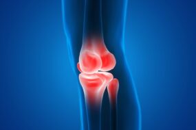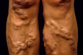
Deep Vein Thrombosis (DVT) is the formation of a blood clot in the deep veins, most commonly in the legs. It poses significant risks, particularly the potential for the thrombus to dislodge and travel to the lungs, leading to a pulmonary embolism (PE).
Pathophysiology
DVT formation is primarily due to Virchow’s triad:
- Stasis of Blood Flow:
- Prolonged immobility (e.g., bed rest, long flights).
- Venous insufficiency or varicose veins.
- Endothelial Injury:
- Trauma (surgery, fractures).
- Inflammation (vasculitis).
- Medical conditions (e.g., atherosclerosis).
- Hypercoagulability:
- Primary Causes: Genetic disorders such as Factor V Leiden, prothrombin mutation, protein C/S deficiency.
- Secondary Causes: Malignancy, pregnancy, hormonal contraceptives, and other medications.
Clinical Presentation
- Symptoms: Often unilateral, including swelling, pain (often described as cramping or heaviness), tenderness, warmth, and discoloration.
- Signs: Swelling in one leg, positive Homan’s sign (calf pain on dorsiflexion), and palpable cord in the area of the thrombus.
Severity Assessment Protocols
To assess the severity of DVT, various scoring systems and clinical assessment tools are used:
- Wells Score:
- A clinical prediction rule used to estimate the probability of DVT.
- Points are assigned based on the following criteria:
- Active cancer (1 point)
- Paralysis, paresis, or recent cast immobilization (1 point)
- Recently bedridden for more than 3 days or major surgery within 12 weeks (1 point)
- Localized tenderness along the distribution of the deep venous system (1 point)
- Swelling of the entire leg (1 point)
- Calf swelling >3 cm compared to the other leg (1 point)
- Pitting edema (1 point)
- Previously documented DVT (1 point)
| Wells Score Interpretation | Risk Category |
| 0 or less | Low risk (1.7% DVT) |
| 1-2 | Moderate risk (16% DVT) |
| 3 or more | High risk (75% DVT) |
Investigation Protocols
- Initial Assessment:
- Clinical Evaluation: History and physical examination focusing on risk factors and symptoms.
- D-Dimer Testing:
- A fibrin degradation product that is elevated in the presence of thrombus.
- Sensitivity is high (up to 95%) but specificity is low, so a positive result requires further investigation.
- Cut-off values: Generally, <250 ng/mL (or age-adjusted thresholds) suggests a low probability of DVT, while higher values necessitate further evaluation.
- Ultrasound:
- Compression Ultrasound: The gold standard for DVT diagnosis, particularly in the calf and thigh veins. If the vein collapses upon compression, it is normal; if it does not, a clot may be present.
- Color Doppler Ultrasound: Can provide additional information on venous blood flow and thrombosis.
- Venography:
- An invasive procedure involving contrast dye injection into a vein, providing detailed images of the venous system. Rarely used due to the availability of non-invasive methods and associated risks.
- Magnetic Resonance Venography (MRV):
- Useful in complicated cases or when the ultrasound findings are inconclusive, especially for pelvic DVT.

Treatment Options
- Anticoagulation Therapy:
- Unfractionated Heparin (UFH):
- Administered IV; initial bolus followed by continuous infusion.
- Dosage:
- Initial: 80 units/kg IV bolus (max 5000 units), followed by 18 units/kg/hr infusion.
- Monitor activated partial thromboplastin time (aPTT) aiming for 1.5-2.5 times the control value.
- Low Molecular Weight Heparin (LMWH) (e.g., Enoxaparin):
- Administered subcutaneously.
- Dosage:
- Initial: 1 mg/kg every 12 hours or 1.5 mg/kg once daily (for outpatient management).
- No routine monitoring required, though anti-factor Xa levels may be monitored in specific populations.
- Warfarin:
- A Vitamin K antagonist used for long-term anticoagulation therapy.
- Mechanism of Action: Warfarin inhibits Vitamin K epoxide reductase, leading to decreased synthesis of Vitamin K-dependent clotting factors (II, VII, IX, X) and proteins C and S.
- INR Monitoring: The International Normalized Ratio (INR) is crucial for monitoring warfarin therapy. The typical therapeutic range for DVT/PE treatment is 2.0 to 3.0.
- Initial Dosing: Usually between 5-10 mg daily, with adjustments based on INR results.
- Monitoring Frequency: Daily or every other day during the initiation phase, then every 1-4 weeks once stable.
- Adjustments:
- INR < 2.0: Increase the dose by 10-20%.
- INR 2.0-3.0: Continue the current dose.
- INR 3.1-4.0: Decrease the dose by 10-20% or hold a dose.
- INR > 4.0: Hold the dose and consider oral Vitamin K (1-2.5 mg) based on bleeding risk.
- Direct Oral Anticoagulants (DOACs):
- Rivaroxaban (Xarelto):
- Dosage: 15 mg twice daily for the first 21 days, followed by 20 mg once daily.
- Apixaban (Eliquis):
- Dosage: 10 mg twice daily for 7 days, then 5 mg twice daily.
- Dabigatran (Pradaxa):
- Requires initial treatment with parenteral anticoagulant for 5-10 days.
- Dosage: 150 mg twice daily thereafter.
- Edoxaban (Savaysa):
- Requires initial treatment with parenteral anticoagulant for 5-10 days.
- Dosage: 60 mg once daily.
- Rivaroxaban (Xarelto):
- Unfractionated Heparin (UFH):
- Thrombolytic Therapy:
- Used in severe DVT or massive PE cases where rapid resolution of the thrombus is critical.
- Common agents: Alteplase, reteplase.
- Dosage:
- Alteplase: 100 mg over 2 hours for DVT.
- Risks include bleeding complications, and this treatment is typically reserved for selected patients.
- Inferior Vena Cava (IVC) Filter:
- Indicated in patients with recurrent DVT/PE despite adequate anticoagulation or those with contraindications to anticoagulants.
- Placed via catheterization, typically in the femoral or jugular vein.
- Compression Therapy:
- Graduated compression stockings (20-30 mmHg) to reduce swelling and promote venous return, often recommended after DVT diagnosis.
Monitoring and Follow-up
- Routine Monitoring:
- Patients on UFH require aPTT monitoring.
- Those on warfarin require INR monitoring.
- For LMWH and DOACs, routine monitoring is not necessary unless indicated.
- Follow-up Ultrasound:
- Recommended in 1-2 weeks to assess clot resolution and rule out complications.
Prevention Strategies
- Mechanical Prophylaxis:
- Use of graduated compression stockings or pneumatic compression devices in high-risk patients.
- Pharmacologic Prophylaxis:
- For hospitalized patients or those undergoing major surgery, LMWH or unfractionated heparin is commonly used.
- Patient Education:
- Encourage mobilization, hydration, and awareness of DVT symptoms, especially during long flights or postoperative recovery.


