- Home
- INTERNAL MEDICINE
- Sickle Cell Disease (SCD)


Sickle cell disease (SCD) is most prevalent in regions where malaria is or was common, such as sub-Saharan Africa, the Mediterranean, the Middle East, and parts of India. Globally, an estimated 300,000 infants are born with SCD each year.
Genetics
SCD is an autosomal recessive disorder caused by a mutation in the β-globin gene (HBB), located on chromosome 11. The mutation results in the substitution of valine for glutamic acid at the sixth position of the β-globin chain, producing hemoglobin S (HbS). When deoxygenated, HbS polymerizes, leading to the sickling of red blood cells (RBCs).
Inheritance Patterns: Individuals with one copy of the HbS gene (heterozygous) have sickle cell trait (SCT), while those with two copies (homozygous) exhibit SCD. Compound heterozygous conditions (e.g., HbSC, HbS/β-thalassemia) also manifest with varying severity of the disease.
Pathophysiology and Molecular Mechanisms
- Polymerization of Deoxygenated HbS:
- The fundamental pathophysiology of SCD lies in the polymerization of deoxygenated HbS, leading to the formation of long fibers that distort RBCs. This polymerization process is dynamic and reversible; cells may return to a biconcave shape upon reoxygenation. However, repeated sickling cycles lead to irreversible damage to the cell membrane and persistent sickling.
- Hemolysis and Nitric Oxide (NO) Dysregulation:
- Hemolysis releases free hemoglobin into the bloodstream, which binds and depletes NO, a potent vasodilator. Reduced NO availability leads to endothelial dysfunction, increased vascular tone, and a pro-thrombotic state, contributing to complications such as pulmonary hypertension and vaso-occlusion.
- Chronic Inflammation and Endothelial Activation:
- Chronic hemolysis and intermittent hypoxia trigger a systemic inflammatory response. Activated endothelial cells express adhesion molecules (e.g., VCAM-1, ICAM-1, P-selectin) that facilitate the adhesion of sickle cells, leukocytes, and platelets to the vascular endothelium, exacerbating vaso-occlusion.
- Oxidative Stress and Red Cell Injury:
- Oxidative damage occurs due to increased production of reactive oxygen species (ROS) and impaired antioxidant defense mechanisms within sickle RBCs. This leads to membrane lipid peroxidation, disruption of membrane proteins, and increased susceptibility to hemolysis.
- Mechanisms of Vaso-Occlusion:
- Vaso-occlusion is a complex process involving the interaction of sickle RBCs with endothelial cells, leukocytes, and platelets. Factors that contribute to vaso-occlusion include:
- Cellular Adhesion: Upregulation of adhesion molecules on the endothelium and RBCs increases the interaction between cells and the vascular wall.
- Altered Blood Flow Dynamics: The presence of rigid sickle cells increases blood viscosity, particularly in post-capillary venules where flow velocity is low, favoring occlusion.
- Inflammatory Mediators: Elevated levels of pro-inflammatory cytokines (e.g., TNF-α, IL-1β, IL-6) and leukotrienes promote endothelial activation and vascular occlusion.
- Vaso-occlusion is a complex process involving the interaction of sickle RBCs with endothelial cells, leukocytes, and platelets. Factors that contribute to vaso-occlusion include:
Clinical Manifestations and Organ-Specific Complications
Vaso-Occlusive Crisis (VOC)
The polymerization of HbS and the resulting sickled shape of RBCs cause obstruction in microvasculature. This leads to ischemia, hypoxia, and severe pain, often triggered by factors such as dehydration, infection, stress, or cold exposure.
VOC can affect any organ system, with common sites being the bones, joints, chest, and abdomen.
- Clinical Presentation:
- Sudden onset of severe pain, typically in the back, legs, arms, abdomen, or chest.
- Accompanied by fever, swelling, or signs of organ dysfunction.
- Management:
- Pain Control:
- Opioids:
- Morphine: 0.05-0.1 mg/kg intravenously (IV) every 2-4 hours or 0.02-0.05 mg/kg/hr continuous infusion. Oral doses of 10-30 mg every 4 hours for adults.
- Hydromorphone: 0.015 mg/kg IV every 3-4 hours. PCA options include 0.2-0.4 mg demand doses with 6-10 minute lockout intervals.
- NSAIDs: To reduce inflammation and opioid requirements.
- Ketorolac: 15-30 mg IV every 6 hours for up to 5 days.
- Ibuprofen: 10 mg/kg orally every 6-8 hours (maximum 2,400 mg daily).
- Opioids:
- Hydration: Administer isotonic fluids at a maintenance rate (1-1.5 times basal) to avoid worsening VOC.
- Oxygen Therapy: For SpO2 < 95% or evidence of hypoxia.
- Hydroxyurea: Long-term therapy starting at 15-20 mg/kg/day, increased to 35 mg/kg/day.
- Hydroxyurea works primarily by increasing fetal hemoglobin (HbF) production, which reduces the concentration of hemoglobin S (HbS) in red blood cells and decreases sickling in sickle cell disease (SCD).
- It inhibits ribonucleotide reductase, thereby reducing DNA synthesis and proliferation of rapidly dividing cells, and also exhibits anti-inflammatory effects by decreasing leukocyte adhesion and migration, which helps prevent vaso-occlusive crises. Additionally, hydroxyurea lowers blood viscosity by increasing plasma volume and reducing the number of sickled cells, enhancing blood flow and oxygen delivery to tissues. Its multifaceted mechanism of action makes it an effective treatment for managing complications of SCD.
- Pain Control:
Acute Chest Syndrome (ACS)
ACS occurs due to vaso-occlusion in pulmonary vasculature, often triggered by infection, fat embolism, or bone marrow embolism. It is characterized by acute respiratory symptoms.
Inflammatory mediators exacerbate the sickling process in pulmonary vessels, leading to hypoxia and acute lung injury.
- Clinical Presentation
- Chest pain, fever, tachypnea, cough, or hypoxia.
- New pulmonary infiltrates seen on chest radiography.
- Management
- Antibiotics: Empiric coverage for typical and atypical organisms.
- Ceftriaxone: 50-75 mg/kg IV every 12-24 hours (maximum 2 g).
- Azithromycin: 10 mg/kg on day 1, followed by 5 mg/kg daily for 4 days.
- Blood Transfusion: To improve oxygenation; aim for HbS < 30%.
- Bronchodilators:
- Albuterol nebulization: 2.5 mg every 4-6 hours.
- Corticosteroids:
- Methylprednisolone: 1-2 mg/kg IV every 12 hours for 2-3 days.
- Antibiotics: Empiric coverage for typical and atypical organisms.
Splenic Sequestration Crisis
Sudden pooling of blood in the spleen leads to rapid-onset splenomegaly and severe anemia. It often occurs in children due to the relatively intact spleen.
- Clinical Presentation:
- Acute abdominal pain, severe anemia, hypovolemic shock, and splenomegaly.
- Management:
- Immediate blood transfusion: PRBCs to restore hemoglobin levels.
- Splenectomy: Consider if recurrent.
- Hydration: Monitor to prevent overhydration.
Aplastic Crisis
Often triggered by infection with parvovirus B19, which suppresses erythropoiesis, leading to sudden severe anemia.
- Clinical Presentation:
- Rapid drop in hemoglobin, fatigue, pallor, and reticulocytopenia.
- Management:
- Blood transfusion: To stabilize hemoglobin.
- Intravenous Immunoglobulin (IVIG): 400 mg/kg for 5 days in immunocompromised individuals.
Hemolytic Crisis
Characterized by acute exacerbation of hemolysis, resulting in a rapid decline in hemoglobin.
- Clinical Presentation:
- Jaundice, dark urine, and worsening anemia.
- Management:
- Hydration: To support kidney function.
- Transfusions: To maintain hemoglobin levels.
- Folic Acid: 1 mg daily to support erythropoiesis.
Priapism
Sickling of RBCs in the penile vasculature leads to prolonged, painful erections due to venous outflow obstruction.
- Clinical Presentation
- Painful erection lasting more than 4 hours.
- Management
- Immediate intervention: Aspiration of blood from the corpus cavernosum.
- Intracavernosal phenylephrine: 100-500 mcg every 3-5 minutes.
- Hydration, pain relief, and potential exchange transfusion for refractory cases.

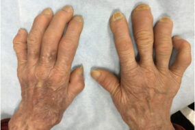
Chronic Complications
- Chronic Pain and Opioid Dependency: Persistent pain due to avascular necrosis (AVN) of bones, chronic inflammation, and neuropathic factors. Opioid use is often necessary for management, but long-term use raises concerns about dependency and tolerance.
- Pulmonary Hypertension: Affects up to 10% of adult SCD patients. Pathogenesis involves hemolysis-induced NO depletion, chronic hypoxia, and endothelial dysfunction. It is associated with high morbidity and mortality.
- Renal Complications: Include hyposthenuria (inability to concentrate urine), hematuria, albuminuria, and progressive glomerulopathy, leading to chronic kidney disease. Sickle cell nephropathy is a common cause of end-stage renal disease in this population.
- Cardiac Issues: Include left ventricular diastolic dysfunction, increased cardiac output due to chronic anemia, and dilated cardiomyopathy. Patients may develop heart failure with preserved or reduced ejection fraction.
- Ocular Complications: Sickle cell retinopathy, which includes proliferative and non-proliferative forms, can lead to retinal detachment and vision loss.
- Hepatobiliary Issues: Include gallstone formation due to chronic hemolysis (leading to bilirubin gallstones), hepatic iron overload from frequent transfusions, and sickle cell intrahepatic cholestasis.
- Leg Ulcers: Often occur on the medial or lateral malleoli, attributed to chronic venous insufficiency and microvascular occlusion. They are difficult to heal and can be recurrent.
Diagnostic Approaches for Sickle Cell Disease (SCD) and Its Complications
Initial Diagnosis of Sickle Cell Disease
- Newborn Screening:
- Most developed countries have implemented newborn screening programs for SCD. The screening is done using:
- High-Performance Liquid Chromatography (HPLC): Identifies the different types of hemoglobin, including hemoglobin S (HbS), hemoglobin A (HbA), and hemoglobin F (HbF).
- Isoelectric Focusing (IEF): Separates hemoglobin variants based on their charge differences. It can distinguish between HbS, HbA, and other abnormal hemoglobins.
- Capillary Electrophoresis (CE): Another method for hemoglobin variant detection.
- DNA Analysis (PCR): Used to confirm mutations in the HBB gene, identifying homozygous (HbSS) or compound heterozygous forms (HbSC, HbSβ-thalassemia).
- Sickle Cell Trait Screening
- Individuals with a positive family history or presenting with symptoms should undergo HPLC, IEF, or DNA testing to confirm if they have sickle cell trait (HbAS) or another hemoglobinopathy.
Diagnostic Tools for Evaluating SCD Complications
- Complete Blood Count (CBC):
- Reveals chronic anemia with hemoglobin levels typically ranging from 6-10 g/dL in homozygous SCD (HbSS).
- Reticulocyte Count: Elevated in response to chronic hemolysis. A decreased reticulocyte count may indicate aplastic crisis.
- Peripheral Blood Smear: Shows sickled cells, target cells, Howell-Jolly bodies (indicative of splenic dysfunction), and nucleated RBCs.
- Hemolysis Markers:
- Lactate Dehydrogenase (LDH), Unconjugated Bilirubin, and Haptoglobin: Elevated LDH and bilirubin levels, and low haptoglobin suggest ongoing hemolysis.
- Iron Studies and Folic Acid Levels: Monitor for iron overload (secondary to transfusions) and folate deficiency due to increased erythropoiesis.
Specific Diagnostic Approaches for Each SCD Crisis
- Vaso-Occlusive Crisis (VOC):
- Clinical Diagnosis: Based on a history of acute pain episodes, particularly in the extremities, chest, or abdomen.
- Imaging (X-rays, MRI): May reveal bone infarction, avascular necrosis, or osteomyelitis if symptoms persist beyond typical pain management.
- Acute Chest Syndrome (ACS):
- Chest X-ray: Detects new pulmonary infiltrates consistent with pneumonia or atelectasis.
- Pulse Oximetry: Evaluates oxygen saturation levels, often decreased in ACS.
- Arterial Blood Gases (ABG): Assesses for hypoxia and acidosis.
- Blood Cultures and Respiratory Pathogen Testing: Helps identify infectious triggers.
- Splenic Sequestration Crisis:
- Physical Examination: Detects splenomegaly, often with acute abdominal pain.
- Ultrasound: Measures spleen size and detects rapid enlargement or infarction.
- CBC: Severe drop in hemoglobin and hematocrit with elevated reticulocyte count.
- Aplastic Crisis:
- CBC: Markedly low hemoglobin levels with a significantly reduced reticulocyte count.
- Parvovirus B19 Serology (IgM and IgG) or PCR Testing: Confirms recent infection.
- Hemolytic Crisis:
- Peripheral Smear and CBC: Reveals increased signs of hemolysis.
- Hemoglobin Electrophoresis: May help identify coexisting hemoglobin variants contributing to hemolysis.
- Priapism:
- Doppler Ultrasound: Assesses blood flow in the penile vasculature.
- CBC and Hemolysis Markers: Helps determine the systemic impact and any ongoing hemolysis.
Additional Diagnostic Tools
- Transcranial Doppler Ultrasound (TCD):
- Used to assess cerebral blood flow velocity in children with SCD. An elevated velocity indicates an increased risk of stroke, guiding the need for prophylactic transfusions.
- Magnetic Resonance Imaging (MRI):
- MRI of the Brain: Detects silent cerebral infarcts and overt strokes.
- MRI of Joints and Bones: Evaluates avascular necrosis, osteomyelitis, or bone infarction.
- Echocardiography:
- Screens for pulmonary hypertension, which is a long-term complication of SCD, by evaluating right ventricular pressure and other hemodynamic parameters.
- Liver Function Tests and Abdominal Imaging:
- Ultrasound or MRI: Monitors for gallstones, liver infarction, and iron overload.
- Bone Marrow Aspiration:
- May be performed in cases of suspected aplastic crisis or unexplained severe anemia to rule out bone marrow suppression.
Sickling Test
The sickling test, also known as the sickle cell preparation test, is a diagnostic tool used to detect the presence of hemoglobin S (HbS), which causes red blood cells to sickle under low oxygen conditions. It falls under screening tests for sickle cell disease and sickle cell trait.
Role and Procedure of the Sickling Test
The sickling test is primarily used as an initial screening tool to detect sickle cell disease (SCD) or sickle cell trait (HbAS). It is less commonly used today because more specific and sensitive tests (like HPLC and hemoglobin electrophoresis) are available.
- Procedure:
- A blood sample is mixed with a deoxygenating agent (such as sodium metabisulfite) to induce hypoxia.
- Under low oxygen conditions, red blood cells containing HbS will sickle and can be seen microscopically.
- Interpretation:
- Positive Test: Presence of sickle-shaped red blood cells indicates that the person has sickle cell trait or disease. However, it does not differentiate between homozygous sickle cell disease (HbSS) and other sickle cell variants (e.g., HbSC, HbSβ-thalassemia).
- Negative Test: Absence of sickled cells typically rules out significant amounts of HbS.
Limitations of the Sickling Test
- Qualitative Rather Than Quantitative: It does not measure the amount of HbS or differentiate between sickle cell disease and sickle cell trait.
- Less Sensitivity: Cannot detect cases with low levels of HbS (e.g., in newborns or after transfusions).
- Modern Replacements: It has largely been replaced by more accurate and specific tests like hemoglobin electrophoresis, high-performance liquid chromatography (HPLC), and DNA testing.

When Is the Sickling Test Used?
- Resource-Limited Settings: It may still be used in places where advanced testing is not readily available.
- Preliminary Screening: In settings where rapid, low-cost screening is needed before confirmatory testing.