- Home
- INTERNAL MEDICINE
- Rheumatology
- Pseudogout


Pseudogout, also known as calcium pyrophosphate deposition disease (CPPD), is a crystal-induced arthritis caused by the deposition of calcium pyrophosphate dihydrate (CPPD) crystals in the joints and surrounding tissues. Unlike gout, which is caused by monosodium urate crystals, pseudogout involves CPPD crystals, leading to joint inflammation and pain.
Pathophysiology
- CPPD crystal formation occurs when calcium and pyrophosphate ions in the synovial fluid combine, leading to crystal deposition in the cartilage (chondrocalcinosis), joint capsules, and periarticular tissues.
- The pathogenesis of CPPD deposition is not fully understood but is thought to involve factors such as:
- Age-related changes: Increased prevalence with aging, suggesting cartilage degradation plays a role.
- Metabolic disorders: Conditions like hyperparathyroidism, hemochromatosis, hypomagnesemia, and hypophosphatasia are associated with increased CPPD deposition.
- Genetic predisposition: Familial cases have been reported, particularly in younger patients with polyarticular involvement.
Clinical Presentation
- Acute CPPD Arthritis (Pseudogout)
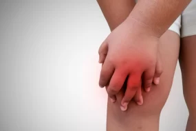
- Characterized by sudden onset of joint pain, swelling, warmth, and redness, resembling acute gout.
- Most commonly affects large joints such as the knees, wrists, shoulders, ankles, and elbows. The first MTP joint (common in gout) is rarely involved.
- Attacks can last days to weeks and may be precipitated by trauma, surgery, or a severe medical illness (e.g., myocardial infarction or stroke).
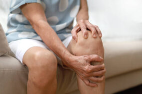
- Chronic CPPD Arthritis
- Mimics osteoarthritis but with atypical features, such as involvement of non-weight-bearing joints (e.g., wrists, shoulders).
- It may present as chronic, low-grade joint pain, morning stiffness, and intermittent flares.
- Asymptomatic Chondrocalcinosis
- Radiographic evidence of CPPD crystal deposition (chondrocalcinosis) may be found in patients without symptoms.
- The knee, wrist, and pubic symphysis are commonly affected sites.
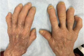
- Pseudo-rheumatoid Arthritis
- May present with polyarticular involvement and symmetric joint distribution, resembling rheumatoid arthritis.
- Pseudo-neuropathic Joint Disease
- Resembles Charcot joint disease, with severe joint destruction and deformity.
Diagnosis
Clinical Evaluation
- Symptoms may resemble other types of arthritis, including gout, rheumatoid arthritis, or osteoarthritis.
- Consider pseudogout in patients with acute monoarthritis or atypical joint distribution.
Laboratory Tests
- Synovial Fluid Analysis: Diagnostic confirmation involves identification of rhomboid-shaped, weakly positively birefringent CPPD crystals under polarized light microscopy.
- Inflammatory Markers: Elevated ESR and CRP may be present during acute attacks.
Imaging
- X-rays: Show chondrocalcinosis, or calcification of the articular cartilage, particularly in the knees, wrists, and symphysis pubis.
- Ultrasound: Can reveal hyperechoic deposits in cartilage and joint effusion.
- CT/MRI: Useful for evaluating atypical joint involvement or spinal pseudogout (CPPD deposition in the cervical spine).
Differential Diagnosis
- Gout: Distinguished by the type of crystal (MSU for gout versus CPPD for pseudogout) and joint involvement.
- Osteoarthritis: Pseudogout may mimic osteoarthritis but involves joints like the wrists and shoulders more frequently.
- Rheumatoid Arthritis: Chronic polyarticular CPPD arthritis can present similarly but without specific rheumatoid factor or anti-CCP antibodies.
Management
Acute CPPD Arthritis (Pseudogout) Treatment
- Nonsteroidal Anti-Inflammatory Drugs (NSAIDs)
- Indomethacin: 50 mg TID for the first 48-72 hours, then taper to 25 mg BID.
- Naproxen: 500 mg BID for 5-7 days, then taper as symptoms resolve.
- Use cautiously in elderly patients or those with renal insufficiency.
- Colchicine
- Useful for acute flare management and prophylaxis.
- Dosage for Acute Attack: 1.2 mg initially, followed by 0.6 mg one hour later. For prophylaxis, 0.6 mg once or twice daily.
- Adjust dose in patients with renal or hepatic impairment.
- Corticosteroids
- Oral prednisone: 30-40 mg daily for 5-7 days, followed by a rapid taper.
- Intra-articular injection: Methylprednisolone (40-80 mg) or triamcinolone (20-40 mg) in larger joints.
- Consider systemic steroids in patients with multiple affected joints or when NSAIDs/colchicine are contraindicated.
- Joint Aspiration and Lavage
- Can provide symptom relief by removing inflammatory mediators and CPPD crystals.
Chronic CPPD Arthritis Management
- Low-Dose Colchicine: 0.6 mg daily to prevent recurrent attacks.
- Physical Therapy: To maintain joint mobility and strength.
- NSAIDs: Used intermittently for flare management, with careful monitoring.
- Address Underlying Conditions: Correct any metabolic abnormalities (e.g., hypomagnesemia, hyperparathyroidism).
Emerging and Experimental Treatments
- IL-1 Inhibitors: Such as anakinra and canakinumab, may benefit refractory cases, though they are not standard treatments.
- Methotrexate and Hydroxychloroquine: Have been used off-label for chronic CPPD arthritis resembling rheumatoid arthritis.
Complications
- Joint Damage and Osteoarthritis: Recurrent inflammation may accelerate joint degeneration.
- Spinal Involvement: CPPD crystals can deposit in the ligamentum flavum, causing crowned dens syndrome (severe neck pain, fever, elevated inflammatory markers).
Monitoring and Follow-Up
- Regular evaluation for joint function and recurrence of acute flares.
- Monitor patients on chronic NSAID or colchicine therapy for potential adverse effects, particularly gastrointestinal or renal complications.