- Home
- INTERNAL MEDICINE
- Rheumatology
- Osteoarthritis


Osteoarthritis is a degenerative joint disorder characterized by the progressive loss of articular cartilage, changes in subchondral bone, synovitis, and osteophyte formation. The pathogenesis involves a complex interplay between mechanical, biochemical, and genetic factors:
- Cartilage Degradation
- In healthy joints, articular cartilage is maintained by a balance between anabolic and catabolic activities of chondrocytes.
- In OA, increased production of matrix-degrading enzymes such as matrix metalloproteinases (MMPs) and aggrecanases (ADAMTS) leads to the breakdown of collagen type II and aggrecan, resulting in cartilage loss.
- Cartilage fragments release into the synovial fluid, promoting synovial inflammation.
- Subchondral Bone Changes
- Increased mechanical loading stimulates bone remodeling, causing subchondral sclerosis and formation of subchondral cysts.
- The resultant bone marrow lesions are associated with pain and joint dysfunction.
- Synovial Inflammation
- Synovitis is present in many OA cases and contributes to symptom severity.
- Inflammatory cytokines (e.g., IL-1β, TNF-α) promote chondrocyte catabolism and synovial inflammation, leading to further cartilage degradation.
- Osteophyte Formation
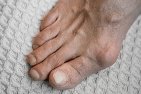
- Growth of new bone at joint margins (osteophytes) occurs in response to mechanical stress and biochemical signaling, leading to joint deformity.
Risk Factors
- Age: Risk increases with advancing age.
- Gender: More common in women, especially after menopause, suggesting hormonal influence.
- Genetics: Family history increases risk, indicating genetic susceptibility.
- Obesity: Excess weight increases joint loading and adipose tissue-derived cytokines contribute to inflammation.
- Joint Injury: Previous joint injuries (e.g., ACL tear, meniscus injury) predispose to OA development.
- Occupation and Physical Activity: Repetitive stress from certain jobs or sports can increase risk.
Clinical Presentation
- Symptoms
- Joint Pain: Worsens with activity and improves with rest; early morning stiffness lasts <30 minutes.
- Joint Stiffness: Particularly after periods of inactivity (gelling phenomenon).
- Limited Range of Motion: Due to pain and structural changes.
- Crepitus: Sensation of grating within the joint.
- Commonly Affected Joints
- Knees: Most commonly affected; weight-bearing stress leads to pain, instability, and deformity (varus or valgus).
- Hips: Causes groin pain radiating to the thigh or buttocks.
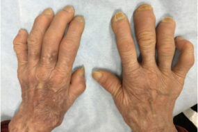
- Hands: Involvement of the distal interphalangeal joints (Heberden’s nodes), proximal interphalangeal joints (Bouchard’s nodes), and first carpometacarpal joint.
- Spine: Involvement of the cervical and lumbar spine may cause radiculopathy.
Diagnosis
- Clinical Evaluation
- Based on history and physical examination findings, including joint pain, stiffness, crepitus, and reduced range of motion.
- American College of Rheumatology (ACR) Criteria for Knee OA Diagnosis:
- Age ≥ 50 years, morning stiffness < 30 minutes, and crepitus on active motion of the knee.
- Radiographic Findings
- Joint Space Narrowing (JSN): Due to cartilage loss.
- Osteophytes: Bony projections at joint margins.
- Subchondral Sclerosis and Cysts: Increased bone density and fluid-filled cysts beneath the cartilage.
- Advanced Imaging
- MRI: Useful for evaluating early cartilage changes, bone marrow lesions, and meniscal tears.
- Ultrasound: Detects synovitis, effusions, and osteophytes.
- Laboratory Tests
- Usually unremarkable; normal ESR and CRP help distinguish OA from inflammatory arthritis (e.g., rheumatoid arthritis).
- Synovial fluid analysis typically shows non-inflammatory characteristics (WBC < 2,000 cells/µL).
Management
1. Non-Pharmacological Management
- Patient Education and Self-Management
- Involves teaching patients about the disease, lifestyle modifications, and coping strategies.
- Weight Loss
- For overweight or obese patients, even a 5% reduction in body weight can significantly alleviate symptoms.
- Exercise
- Low-impact aerobic activities (e.g., swimming, cycling) and strengthening exercises for quadriceps and hamstrings improve joint stability and function.
- Physical Therapy
- Focus on range of motion, muscle strengthening, and joint stabilization.
- Assistive Devices
- Use of braces, orthotics, or canes to reduce joint loading.
2. Pharmacological Management
- Analgesics and NSAIDs
- Acetaminophen: Initial choice for mild pain; up to 3-4 g/day, with caution in liver disease.
- NSAIDs: More effective for moderate to severe pain.
- Ibuprofen: 400-800 mg TID-QID.
- Naproxen: 500 mg BID.
- Celecoxib (COX-2 inhibitor): 100-200 mg once or twice daily; preferred in patients at risk for gastrointestinal complications.
- Topical NSAIDs: Diclofenac gel can be applied to joints (e.g., 4 g QID for knees, hands).
- Intra-Articular Injections
- Corticosteroids: Triamcinolone acetonide (40 mg for large joints) every 3-4 months for acute exacerbations.
- Hyaluronic Acid: Viscosupplementation to improve joint lubrication, although evidence of efficacy is variable.
- Other Medications
- Duloxetine: Approved for chronic musculoskeletal pain, 30-60 mg daily.
- Tramadol: For patients with inadequate response to NSAIDs; start at 25 mg and titrate to 50-100 mg every 4-6 hours.
3. Surgical Management
- Arthroscopy: Indicated for mechanical symptoms (locking, catching) due to loose bodies.
- Osteotomy: Realignment procedures for unicompartmental knee OA.
- Joint Replacement (Arthroplasty)
- Total Knee Arthroplasty (TKA) or Total Hip Arthroplasty (THA): For patients with severe pain and functional limitations refractory to conservative measures.
- Unicompartmental Knee Arthroplasty: Suitable for isolated medial or lateral compartment knee OA.
Complications
- Progressive Joint Damage: May lead to significant disability.
- Spinal OA: Can cause nerve impingement, spinal stenosis, and radiculopathy.
- Secondary Infections: Following intra-articular injections or surgical interventions.
- Comorbidities: Obesity, cardiovascular disease, and depression are commonly associated.
Monitoring and Follow-Up
- Regular Assessment: Evaluate pain, joint function, and side effects of ongoing treatment.
- Radiographic Monitoring: To assess joint deterioration and guide the need for surgical intervention.
- Adjust Management Based on Disease Progression: Escalate treatment for worsening symptoms.
Emerging Treatments and Future Directions
- Disease-Modifying Osteoarthritis Drugs (DMOADs): Potential agents targeting cartilage regeneration, including anti-NGF antibodies, matrix metalloproteinase inhibitors, and intra-articular gene therapy.
- Platelet-Rich Plasma (PRP): Intra-articular injections for symptom relief, though more research is needed.
- Stem Cell Therapy: Being explored for regenerative potential in damaged cartilage.