- Home
- INTERNAL MEDICINE
- Rheumatology
- Juvenile Idiopathic Arthritis ...


Juvenile Idiopathic Arthritis (JIA) is the most common chronic rheumatic disease in children, characterized by persistent arthritis for at least six weeks in a child younger than 16 years, with no identifiable cause. JIA encompasses a group of disorders that vary in presentation, disease course, and prognosis.
Pathophysiology
- Autoimmune Mechanism
- JIA is believed to result from a combination of genetic susceptibility and environmental triggers (e.g., infections) that lead to an autoimmune response targeting synovial tissue.
- The exact mechanism remains unknown, but T-cell activation and pro-inflammatory cytokine production (e.g., TNF-α, IL-1, IL-6) play critical roles in sustaining synovial inflammation.
- Synovial inflammation leads to synovial hyperplasia, increased vascularity, and pannus formation, which can erode cartilage and bone.
- Genetic Factors
- Several genetic associations have been identified, including:
- HLA associations: Particularly HLA-DRB1, which may influence susceptibility to different JIA subtypes.
- Non-HLA genes: PTPN22 and STAT4 have been linked to autoimmune disorders, including JIA.
- Several genetic associations have been identified, including:
- Environmental Triggers
- Infections may act as triggers in genetically predisposed individuals by mimicking host antigens or directly activating the immune system.
Classification
JIA is classified into several subtypes based on the International League of Associations for Rheumatology (ILAR) criteria:
- Oligoarticular JIA
- Involves ≤4 joints during the first six months of disease.
- The most common subtype, accounting for 50-60% of cases.
- Persistent Oligoarticular JIA: Remains limited to ≤4 joints.
- Extended Oligoarticular JIA: Involves more than four joints after the first six months.
- Complications: Uveitis is common, especially in young girls with a positive antinuclear antibody (ANA).
- Polyarticular JIA (RF-negative and RF-positive)
- Involves ≥5 joints within the first six months of disease.
- RF-negative Polyarticular JIA: Often affects smaller joints (e.g., wrists, hands) and is more common in younger children.
- RF-positive Polyarticular JIA: Similar to adult rheumatoid arthritis and is more aggressive, usually presenting in adolescence.
- Systemic JIA (sJIA)
- Characterized by arthritis accompanied by systemic features, such as quotidian fever, salmon-pink macular rash, hepatosplenomegaly, lymphadenopathy, and serositis.
- Accounts for about 10% of JIA cases.
- Pathophysiology involves excessive production of IL-1 and IL-6, which drives systemic inflammation.
- Enthesitis-Related Arthritis (ERA)
- Includes arthritis and enthesitis (inflammation at tendon insertion sites).
- Predominantly affects males over the age of six.
- Often associated with HLA-B27, increasing the risk for developing ankylosing spondylitis.
- Psoriatic Arthritis
- Characterized by arthritis associated with psoriasis or a family history of psoriasis.
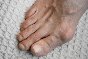
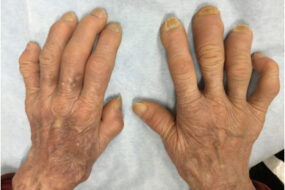
- Patients may present with dactylitis (“sausage digits”) or nail pitting.
- Undifferentiated Arthritis
- Refers to arthritis that does not fit into the above categories or fits into more than one category.
Clinical Presentation
- General Symptoms
- Joint Pain and Swelling: Pain is typically worse in the morning (morning stiffness) and improves throughout the day.
- Joint Warmth and Redness: More common in larger joints (knees, ankles).
- Growth Abnormalities: Chronic inflammation may cause accelerated growth in the affected limb (due to increased blood flow) or growth retardation due to long-term corticosteroid use.
- Subtype-Specific Symptoms
- Systemic JIA: Features daily fevers, evanescent rash, and may involve systemic symptoms like serositis (pericarditis, pleuritis).
- Oligoarticular JIA: Predominantly affects large joints (knees, ankles) and carries a high risk of chronic uveitis.
- Polyarticular JIA: Resembles adult rheumatoid arthritis, especially the RF-positive type, affecting multiple joints symmetrically.
- Enthesitis-Related Arthritis (ERA): Commonly involves the lower limbs (hips, knees, ankles) and may present with inflammatory back pain.
Diagnosis
- Clinical Evaluation
- JIA is primarily a clinical diagnosis based on the presence of persistent arthritis for ≥6 weeks in a child <16 years of age, after excluding other causes (e.g., infections, trauma, malignancy).
- Laboratory Tests
- Complete Blood Count (CBC): May show anemia of chronic disease, leukocytosis, or thrombocytosis, especially in systemic JIA.
- Inflammatory Markers: Elevated ESR and CRP levels indicate active inflammation.
- Autoantibodies:
- ANA: Commonly positive in oligoarticular JIA and associated with uveitis risk.
- Rheumatoid Factor (RF): Positive in RF-positive polyarticular JIA.
- Anti-Cyclic Citrullinated Peptide (Anti-CCP): May be positive in RF-positive polyarticular JIA.
- HLA-B27 Testing: Can support the diagnosis of ERA.
- Imaging
- X-rays: Early stages may show soft tissue swelling; advanced cases can reveal joint space narrowing and bone erosions.
- MRI: Preferred for detecting early joint changes, synovitis, and tenosynovitis.
- Ultrasound: Useful for detecting synovial inflammation and effusion.
- Slit-Lamp Examination
- Regular screening for uveitis is crucial, particularly in ANA-positive oligoarticular JIA.
Management
- Non-Pharmacologic Therapy
- Physical Therapy and Occupational Therapy: Maintain joint function and muscle strength.
- Exercise Programs: Low-impact activities (e.g., swimming, cycling) are encouraged.
- Psychosocial Support: Essential for coping with chronic illness.
- Pharmacologic Therapy
- Non-Steroidal Anti-Inflammatory Drugs (NSAIDs)
- Provide symptomatic relief for pain and inflammation.
- Dosing: E.g., Ibuprofen 30-50 mg/kg/day divided every 6-8 hours.
- Disease-Modifying Anti-Rheumatic Drugs (DMARDs)
- Methotrexate is the first-line DMARD for JIA, particularly polyarticular or systemic forms.
- Dosing: Methotrexate 10-15 mg/m²/week subcutaneously or orally.
- Biologic Agents
- Target specific cytokines involved in JIA pathogenesis:
- TNF-α Inhibitors (e.g., etanercept, adalimumab, infliximab): Effective for polyarticular JIA and ERA.
- IL-1 Inhibitors (e.g., anakinra) and IL-6 Inhibitors (e.g., tocilizumab) are useful for systemic JIA.
- Abatacept (T-cell costimulation modulator) for cases refractory to TNF inhibitors.
- Target specific cytokines involved in JIA pathogenesis:
- Corticosteroids
- Used for systemic symptoms or severe flares, but limited due to side effects.
- Intra-articular triamcinolone injections may be beneficial for oligoarticular JIA.
- Topical Treatments
- Mydriatic eye drops for uveitis prevention in oligoarticular JIA.
- Non-Steroidal Anti-Inflammatory Drugs (NSAIDs)
- Surgical Interventions
- Synovectomy or joint replacement may be necessary for severe joint damage or growth disturbances.
Prognosis
- Outcomes Vary by Subtype
- Oligoarticular JIA: Often has a good prognosis but requires vigilance for uveitis.
- Polyarticular JIA (RF-positive): More likely to have persistent active disease and joint damage.
- Systemic JIA: Can lead to macrophage activation syndrome (MAS), a life-threatening complication.
- Factors Influencing Prognosis
- Early treatment initiation improves long-term outcomes.
- ANA positivity in oligoarticular JIA increases the risk for chronic uveitis.
- Complications
- Growth disturbances (e.g., leg length discrepancy, growth retardation).
- Chronic uveitis, especially in oligoarticular and polyarticular JIA.