Bronchiectasis refers to an abnormal widening of the bronchi. It is a chronic airway disease that shares many features with COPD. It may have one or more exacerbations per year, and the prevalence increases with age.
Etiology
- Congenital or acquired
1. Congenital causes;
- Cystic fibrosis
- Primary ciliary dyskinesia
- Kartagener’s syndrome
- Primary hypogammaglobulinemia
- Young syndrome
2. Acquired causes;
- Pulmonary infections- tuberculosis and pneumonia
- Severe infections in infancy and childhood- whooping cough, measles
- Asthma
- Bronchial tumors
- Cigarette smoking and COPD
- Rheumatic diseases
- Allergic bronchopulmonary aspergillosis
- Airway obstruction- e.g., foreign body aspiration
- Inhaled toxins
Pathophysiology
Two factors must be present;
1. Infectious process
2. Poor clearance of mucus secretions- airway obstruction, defect in host defense, ciliary dysfunction, etc
These cause inflammation, edema, ulceration, and neovascularisation in the airways. This damages the airways leading to dilatation and chronic inflammation. Fibrotic changes occur in the normal surrounding tissue, leading to progressive destruction of lung architecture in advanced disease
Clinical presentation
- Chronic cough, on most days of the week
- Mucopurulent and tenacious sputum on most days of the week
- History of exacerbations- characterized by increased sputum, fever, anorexia, and malaise
- Pleuritic pain
- Hemoptysis- streaks of blood, larger volumes with exacerbations
- Dyspnoea
Physical examination– wheezing, crackles, diminished breath sounds with a blocked proximal bronchus.
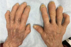
- Digital clubbing in some patients
- Bronchial breathing when there is scarring
- Others- fatigue, anosmia, general debility, halitosis
Investigations
- Complete blood count- leucocytosis. Thrombocytosis is associated with increased mortality.
- Serology- quantification of IgG, IgM, and IgA
- Testing for cystic fibrosis-sweat chloride test or mutational analysis
- Sputum smear for microscopy, culture, and sensitivity
- Chest x-ray- usually not apparent. Thickened airway walls and dilated airways( tram or parallel lines), irregular opacities, areas of consolation or collapse
- CT chest- MDCT( multi-detector computed tomography) is preferred. Reliable CT signs include;
- Tram truck appearance- lack of tapering of bronchi
- Visibility of an airway with 1cm of a costal pleural surface or touching the pleura
- Airway to an arterial ratio of ≥1.5
- Lung function tests- obstructive findings. A very low FVC in advanced disease
Differential diagnosis
- COPD
- Chronic bronchitis
- Asthma
- Tuberculosis
- Pneumonia
- Pulmonary embolism
Management
Acute exacerbation
1. Airflow obstruction- inhaled bronchodilators and glucocorticoids
- Mucoactive agents
2. Oxygen therapy
3. Antibiotics
A) oral- for clinically stable. Treatment for 10 to 14 days
- If culture results are not available or empiric treatment- broad-spectrum fluoroquinolone, e.g., levofloxacin, moxifloxacin
- Sensitive organisms- amoxicillin 500mg TDS
- Beta lactamase positive- amoxicillin-clavulanate
- Pseudomonas- ciprofloxacin
B ) intravenous-severe disease
Antiviral therapy for those exacerbations triggered by the influenza virus
Management of hemoptysis;
- Resuscitate the patient first
- Bronchoscopic techniques- balloon tamponade, laser therapy, cryotherapy, topical vasoconstrictor
- Arteriographic embolization
Management in the long term
- Advise patient to stop smoking
- Physiotherapy- train patient on mechanisms to assist with drainage of excess bronchial secretions
- Manage underlying cause
- Surgical resection- in a small number of patients whose disease is confined to one lobe
- Vaccination for influenza and pneumococcal pneumonia
- Lung transplant
Prognosis
- The disease is progressive when associated with ciliary dysfunction
- It can be good in some patients with aggressive use of antibiotics and regular physiotherapy
Prevention
- Early recognition and treatment of bronchial obstruction
- Prophylaxis and prompt treatment of whooping cough or primary tuberculosis in childhood


