- Home
- GYNECOLOGY/OBSTETRICS
- Molar Pregnancy


Molar pregnancy, or hydatidiform mole, is a type of gestational trophoblastic disease characterized by the abnormal growth of placental tissue. It arises from the fertilization of an abnormal egg, leading to excessive proliferation of trophoblastic cells, which can cause complications ranging from benign to malignant forms of disease.
Types of Molar Pregnancy
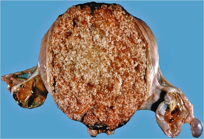
1. Complete Molar Pregnancy
- Definition: A complete mole occurs when an empty egg (ovum) is fertilized by a sperm. The sperm’s genetic material duplicates, resulting in a 46,XX karyotype. There is no fetal tissue.
- Characteristics:
- The chorionic villi are hydropic (swollen) and appear as clusters of cysts resembling grapes.
- The uterine size is often larger than expected for the gestational age.
- No identifiable fetal structures or heart activity.
- Clinical Course: Complete moles are more likely to progress to persistent gestational trophoblastic disease (GTD) or choriocarcinoma compared to partial moles. They can result in significant maternal morbidity and require careful monitoring.
2. Partial Molar Pregnancy
- Definition: A partial mole occurs when a normal egg is fertilized by two sperm, resulting in a triploid karyotype (69 chromosomes, such as 69,XXX or 69,XXY).
- Characteristics:
- There may be some normal fetal tissue, but it is usually malformed and non-viable.
- The placental tissue has a mix of normal and abnormal chorionic villi, with some cystic changes.
- Clinical Course: Partial moles have a lower risk of developing into malignant GTD compared to complete moles, but they still require monitoring for potential complications.
Pathophysiology
- Fertilization: Molar pregnancies result from abnormal fertilization processes. In complete moles, the egg is either fertilized by a sperm that duplicates itself or two sperm simultaneously fertilizing the egg. In partial moles, two sperm fertilize a normal egg.
- Trophoblastic Proliferation: The trophoblasts (the outer layer of the blastocyst) proliferate excessively, leading to abnormal placental tissue growth. This overgrowth can interfere with normal placental function and hormone production.
- Beta-hCG Levels: The abnormal tissue secretes high levels of human chorionic gonadotropin (hCG), often resulting in hyperemesis gravidarum and other complications.
Epidemiology
- Incidence: Molar pregnancies are relatively rare, occurring in about 1 in 1,000 pregnancies in the United States. The incidence is higher in certain populations, such as women of Asian descent and those living in areas with nutritional deficiencies.
- Maternal Age: The risk is higher in women under 20 or over 35 years of age.
Risk Factors
- History of Molar Pregnancy: Women who have previously experienced a molar pregnancy are at higher risk for subsequent occurrences.
- Dietary Factors: Nutritional deficiencies, particularly low levels of carotene and folate, may be linked to increased risk.
- Maternal Conditions: Certain pre-existing maternal health conditions and reproductive history can contribute to the risk.
Clinical Presentation
- Vaginal Bleeding: The most common presenting symptom, typically in the first trimester. The bleeding may be bright red or dark brown.
- Excessive Nausea and Vomiting: Due to markedly elevated hCG levels, leading to hyperemesis gravidarum in some cases.
- Uterine Size Greater Than Expected: The uterus may be larger than the gestational age suggests, often noted during routine examinations.
- Passage of Grapelike Vesicles: In cases of complete moles, women may pass cystic structures resembling grapes.
- Preeclampsia Before 20 Weeks: This is atypical for normal pregnancies and may indicate molar pregnancy.
- Hyperthyroidism Symptoms: Elevated hCG can lead to symptoms like palpitations, tremors, and heat intolerance due to cross-reactivity with thyroid receptors.
Diagnosis
- Ultrasound:
- Transvaginal Ultrasound: The gold standard for diagnosis.
- Complete Mole: Characterized by the “snowstorm” appearance, with numerous cystic spaces and no identifiable fetal structures.
- Partial Mole: May show an abnormal placenta with cystic areas and some evidence of malformed fetal tissue.
- Serum Beta-hCG:
- Levels are significantly elevated in complete moles, often exceeding 100,000 mIU/mL.
- In partial moles, beta-hCG levels are elevated but not as high as in complete moles.
- Histopathological Examination:
- Tissue obtained from evacuation is examined to confirm the diagnosis.
- Features include hydropic degeneration of villi, trophoblastic hyperplasia, and absence of normal fetal tissue in complete moles.
Differential Diagnosis
- Spontaneous Abortion: Early pregnancy loss with bleeding and cramping.
- Ectopic Pregnancy: May present with similar symptoms, but typically with unilateral pelvic pain.
- Choriocarcinoma: A malignant form of GTD that can occur after a molar pregnancy.
- Gestational Trophoblastic Neoplasia: Other forms of trophoblastic disease that may have similar presentations.
Management
- Surgical Management:
- Evacuation of the Uterus: The primary treatment method.
- Suction Curettage: Performed under ultrasound guidance to ensure complete removal of molar tissue.
- D&C (Dilation and Curettage): May also be performed, but suction curettage is preferred for complete evacuation.
- Hysterectomy: Considered in women who are not interested in preserving fertility or in cases of complications.
- Evacuation of the Uterus: The primary treatment method.
- Post-Evacuation Monitoring:
- Serial Beta-hCG Measurements: Monitored weekly until levels become undetectable, then monthly for 6 months.
- Persistent elevated or rising beta-hCG levels indicate the potential for persistent GTD and require further management.
- Follow-Up Care:
- Regular follow-up is essential to monitor for any signs of persistent disease or malignant transformation.
- Education on the importance of monitoring and potential symptoms to watch for is crucial.
- Contraception:
- Effective contraception is recommended for at least 6-12 months post-treatment to avoid confusion between a new pregnancy and residual molar tissue.
Complications
- Persistent Gestational Trophoblastic Disease (GTD):
- Occurs in approximately 15-20% of complete moles and 2-4% of partial moles.
- Requires further evaluation and treatment with chemotherapy in some cases.
- Choriocarcinoma:
- A malignant form of GTD that can develop from either type of molar pregnancy.
- Metastasis can occur to distant sites, such as the lungs and liver.
- Heavy Bleeding: Due to the vascularity of the molar tissue and complications during surgical evacuation.
- Uterine Perforation: Can occur during curettage, especially in cases of very enlarged uteri.
Prognosis
- Complete Mole: Good prognosis with appropriate management; approximately 80% of cases resolve with evacuation. However, there is a significant risk of persistent GTD in about 15-20%.
- Partial Mole: Generally favorable outcomes; lower rates of malignant transformation.
Prevention and Counseling
- Genetic Counseling: Recommended for women with a history of recurrent molar pregnancies.
- Future Pregnancies: Women are advised to have early ultrasound evaluations in subsequent pregnancies to monitor for potential recurrence.
Conclusion
Molar pregnancy is a unique form of gestational trophoblastic disease that requires early recognition and management to prevent complications. With prompt diagnosis and appropriate treatment, the prognosis for women with molar pregnancy is generally favorable, but careful follow-up is essential to monitor for persistent disease and manage any potential complications.


