Shock is a life-threatening condition due to circulatory failure, characterized by inadequate oxygen supply to tissues, producing tissue hypoxia and cellular dysfunction and, ultimately, irreversible tissue damage and death.
Prompt diagnosis and treatment are critical in preventing multi-organ failure(MOF) and death.
Types of shock
1. Distributive shock– characterized by severe peripheral vasodilatation
a) Septic shock- the most common cause of distributive shock. It is a subset of sepsis and can be identified by elevated lactate and the use of vasopressors
b) Neurogenic shock- found in a severe traumatic brain or spinal cord injury. It is caused by autonomic failure, causing low vascular resistance and altered vagal tone
c) Anaphylactic shock- encountered in patients with severe IgE mediated allergic reactions
d) Others- drug or toxin-induced shock, endocrine shock
2. Cardiogenic shock– due to intracardiac causes resulting in reduced cardiac output. The causes include;
- Arrhythmias
- Cardiomyopathy,
- Ischemia
- Mechanical, e.g., Valvulopathies
Two types;
a) Dry and cold – no evidence of congestion
b) Wet and dry- evidence of congestion
3. Hypovolemic shock
a) Hemorrhagic- trauma, upper or lower GI bleeding, perioperative hemorrhage, ruptured aneurysm, tumors, iatrogenic, e.g., biopsy, postpartum hemorrhage, ruptured hematomas, etc
b) Nonhemorrhagic-
- GI losses- vomiting and diarrhea.
- Skin losses- burns, SJS, heat stroke
- Renal losses- osmotic or drug-induced diuresis, salt wasting nephropathies
- Third space losses into body cavities or the extravascular space- post-operative, pancreatitis
Classification of hemorrhagic shock;
I. <15% or <750 ml blood loss
II. 15 to 30% or 750 to 1500ml blood loss
III. 30-40% or 1500 to 2000ml blood loss
IV. >40% or >2000ml blood loss
4) Obstructive shock– due to extracardiac causes of cardiac pump failure
a) pulmonary vascular- pulmonary embolism, severe pulmonary hypertension
b) mechanical causes- constrictive pericarditis, tension pneumothorax, pericardial tamponade, restrictive cardiomyopathy
*Patients can have combined forms of shock
Stages of shock
1. Pre shock;
- Also known as compensated shock.
- Characterized by compensatory mechanisms to reduced tissue perfusion. E.g., tachycardia, peripheral vasoconstriction,
- Clinical presentation- tachycardia, modest reduction in the systolic blood pressure, or a mild to moderate hyperlactatemia may be the only clinical signs
- Deterioration can be prevented
2. Shock;
- Compensatory mechanisms are overwhelmed, and signs of organ dysfunction appear; symptomatic tachycardia, hypotension, dyspnea, metabolic acidosis, oliguria, diaphoresis, and cold, clammy skin
- In hypovolemic shock, signs and symptoms occur with a blood loss of 20 to 25% of the blood volume, while in cardiogenic shock, a fall in the cardiac index to less than 2.5 L/min/m2
3. End organ dysfunction;
- The progressive shock will lead to irreversible tissue damage, MOF, and death.
- It is characterized by anuria, acute renal failure, further reduction in cardiac output, severe hypotension, severe hyperlactatemia, and coma.
- Death is common in this stage
* Hypotension may be absent in some patients
* Signs of heart failure in shock are signs of cardiogenic shock
Initial management
- Start with the ABCDEs
- Conditions needing lifesaving interventions- tension pneumothorax, cardiac tamponade, anaphylactic shock, significant hemorrhage, arrhythmias, septic shock, aortic dissection, adrenal crisis, animal bites, etc
- Gain intravenous access through 2 wide bore cannulas. Draw blood for grouping and cross-matching. A central venous line may be inserted.
- Start cardiac monitoring- pulse oximetry, central venous pressure,
- If possible, classify the shock and determine the cause
- Begin intravenous fluids resuscitation if there are no signs of overload.
- Determine the need for blood transfusion and the use of vasopressors and inotropes. The first line vasopressor in undifferentiated shock is norepinephrine.
- Call the rapid response team.
Initial diagnostic investigations;
Laboratory-
- Serum lactate,
- CBC,
- A complete metabolic panel,
- Cardiac enzymes and natriuretic peptides
- Coagulation studies
- D Dimer level
- Blood gas analysis
- ECG
- Bloods glucose
Imaging;
- Chest Xray
- Others based on the etiology
Further tests;
1. Hemorrhagic shock;
- FAST scan
- DRE for GI bleeding
- CT abdomen for abdominal losses
- Renal function tests
2. Cardiogenic shock–
- Echocardiogram,
- BNP and NT-proBNP
3. Obstructive shock;
- Echocardiogram and POCUS
- CTPA
4. Distributive shock
- Septic shock- acute phase reactants, Cultures, Urinalysis
- Anaphylactic shock- serum mast cell tryptase
- Neurogenic shock- CT or MRI of the brain or spinal cord
Definitive management
1. Hypovolemic shock
A) Hemorrhagic-
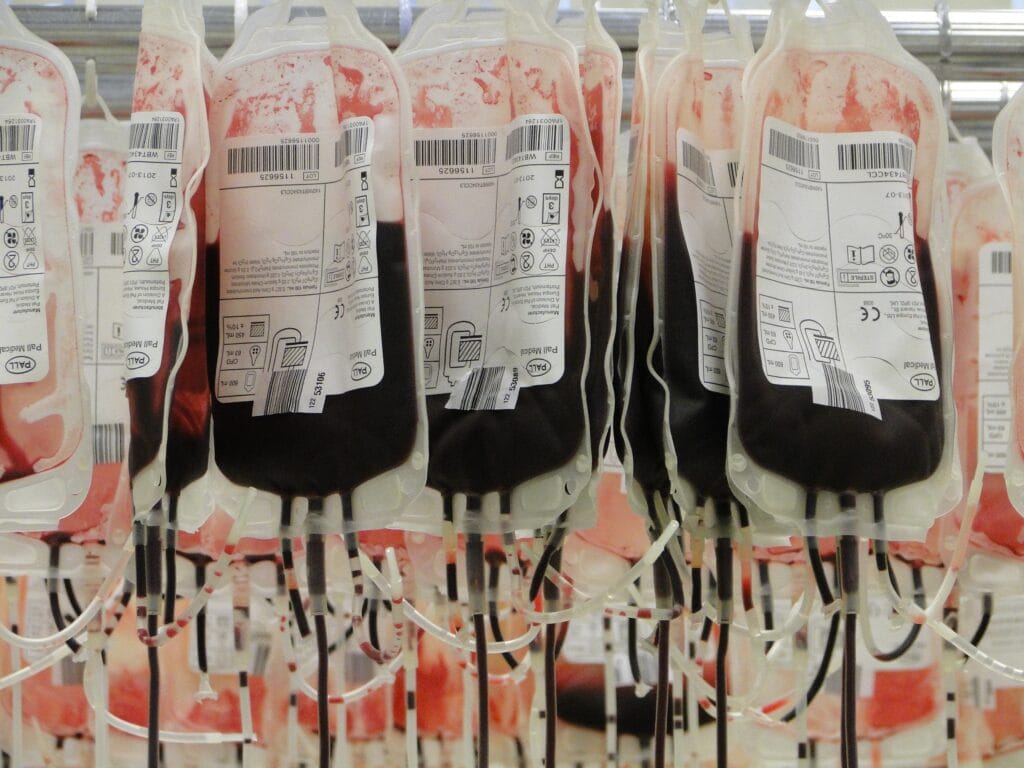
- Blood and blood products- initially transfused with group O blood. Switch to cross-matched blood as soon as possible. Give O negative blood to women of childbearing age.
- In a massive transfusion, transfuse RBC, platelets, and plasma at the ratio of 1:1:1
- Apply pressure to bleeding wounds, suture or staple open wounds
- Consult the relevant specialist based on the etiology
B) Nonhemorrhagic-
Manage the underlying cause-
- Antiemetics for vomiting
- Cessation of diuretics and management of diabetes insipidus for renal losses
- Management of burns
- Management of pancreatitis and bowel obstruction
- Replace ongoing fluid losses
- Treat electrolyte abnormalities
2. Cardiogenic shock-
a) Wet and cold-
- Administer inotropes, e.g., dobutamine, dopamine, milrinone
- If shock persists, give a vasopressor- norepinephrine
- If systolic blood pressure is > 90 mmHg, start diuretics
- If symptoms persist, treat as refractory acute heart failure
b). Dry and cold-
- Start with a fluid bolus
- Consider a fluid challenge
- If symptoms persist, give a vasopressor
- Administer inotropes if shock persists
3. Obstructive shock-
- Consider vasopressors or inotropes
- Treat According to etiology;
- Pericardiocentesis for cardiac tamponade
- Needle thoracostomy followed by chest tube for tension pneumothorax
- Thrombolytic therapy for PE
4. Distributive shock
- Reverse vasodilatation with vasopressors
A) Septic shock-
- Give corticosteroids if the shock is resistant to the first vasopressor. Dopamine and dobutamine are additional options
- Lactate guided fluid resuscitation
- Broad-spectrum antibiotics
- The mean arterial pressure target is ≥65 mmhg
B) Anaphylactic shock-
- Secure the airway and remove allergen if possible
- IM epinephrine 1:1000 as needed
- If refractory, start epinephrine IV 1:1000000
- If still refractory, consider other vasopressors, give glucagon if on a beta-blocker, and ensure an adequate fluid status
C) Neurogenic shock-
- Give vasopressors
- Consider atropine for bradycardia
- Consult with a spine specialist
- Monitor urine output
- Monitor for any cardiovascular complications
Complications of shock;
- Cardiogenic shock- pulmonary edema
- Anaphylactic shock- airway obstruction
- Hypovolemic and septic shock- DIC
- General- acute renal failure, delirium
- Nosocomial infections
- Fluid overload
- Complications of mechanical ventilation


