

Pneumothorax is defined as gas in the pleural space
Aetiology
1. Primary spontaneous pneumothorax( PSP)
-Presents without a precipitating external event in the absence of clinical lung disease
Risk factors-
- Smoking- airway inflammation and respiratory bronchiolitis
- Subpleural blebs- the mechanism is not well known.
- Genetic predisposition- reports have shown clustering of PSP in families
- Male sex,
- Slim, tall stature
- Homocystinuria
- A drop in atmospheric pressure
2. Secondary spontaneous pneumothorax ( SSP)
Pneumothorax presents as a complication of underlying lung disease
Causes;
- COPD
- Necrotizing lung infections- TB, pneumocystis pneumonia, SARS- CoV-2
- Malignancy
- Cystic fibrosis
- Sarcoidosis
- Cystic lung diseases, e.g. Sjogren syndrome
- Catamenial pneumothorax
- HIV
- Abnormalities of the pleural membrane, e.g. Marfan Syndrome
3. Traumatic pneumothorax– blunt or penetrating trauma. It is probably the most common cause of pneumothorax
4. Miscellaneous– exercise, illicit drugs, scuba diving, air travel, immunosuppressant drugs, anorexia nervosa
*Tension pneumothorax– a type of pneumothorax characterised by a progressive increase in pressure in the thoracic cavity and cardiorespiratory compromise
Clinical presentation
- Ranges from asymptomatic to having features of cardiorespiratory compromise
- Most often presents with sudden onset dyspnoea and pleuritic chest pain. The pain is usually unilateral but can be central or bilateral.
Physical examination;
- in a small pneumothorax, findings may be restricted to those of the underlying lung disease.
- Large pneumothorax- decreased chest expansion on the affected side, reduced or absent breath sounds, absent tactile or vocal fremitus, hyper resonant percussion, subcutaneous emphysema.
- Evidence of laboured breathing
Tension pneumothorax-
- acute severe respiratory distress
- Cyanosis, diaphoresis and restlessness
- Distended neck veins and hemodynamic instability
- Reduced chest expansion on the ipsilateral side
- Tracheal deviation- a late sign
- Presence of lacerations or stab wound
- Tachycardia, hypotension, rapid decrease in SPO2
Laboratory findings
-Non-specific
- May show mild leucocytosis
- Helpful in identifying underlying diseases, e.g. D dimer, troponins to identify the cause of chest pain
- Arterial blood gases- obtained depending on the clinical status of the patient
- ECG findings are non-specific. May reveal a sinus tachycardia
Imaging
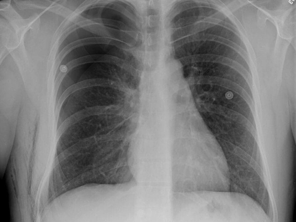
1. Chest Xray-
- Simple pneumothorax- gas in the pleural space with no mediastinal shift. Patients are clinically stable
- Tension pneumothorax- mediastinal shift, splaying of the ribs, flattening of ipsilateral diaphragm
2. Pleural ultrasonography- in unstable patients
3. Chest CT- best modality for determining size and location
Other tests- lung function tests, specific aetiology tests
Management
- Assess for stability- ABCDEs
- Provide respiratory support
- Assess the type and severity of the pneumothorax
- Tension pneumothorax and open pneumothorax are life-threatening conditions that require immediate intervention.
- Unstable or high-risk patients- emergent chest decompression
- Spontaneous pneumothorax- for low risk, treat conservatively; for high risk, put a chest tube
- Traumatic pneumothorax- most will require a chest tube
- Manage the underlying cause
Definitive measures to prevent a recurrence
Surgery-
- Pleurodesis
- Bleb closure
- Lung volume reduction
Non-surgical candidates- chemical pleurodesis with tetracycline derivatives, talc and others


